The given diagram shows a stage during mitotic division in an animal cell: The given diagram shows a stage during mitotic division in an animal cell:
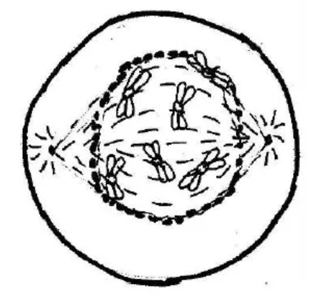
Draw a neat labelled diagram of the cell as it would appear in the next stage. Name the stage.
Topic: Metaphase — diagram
Answer
The next stage is METAPHASE. Your diagram must show: • the chromosomes arranged at the EQUATOR of the cell, in a single plane • each chromosome still DOUBLE — two CHROMATIDS joined at a CENTROMERE • SPINDLE FIBRES running from the poles and ATTACHED to the centromeres • two CENTRIOLES with ASTERS at opposite poles (this is an ANIMAL cell) • NO nuclear membrane and NO nucleolus • CELL MEMBRANE only — no cell wall
Labels required: chromosome, chromatid, centromere, spindle fibre, centriole, aster, equator, cell membrane.
WHAT IDENTIFIES METAPHASE, and what the examiner looks for first: the chromosomes LINED UP AT THE EQUATOR in one plane, with the spindle fibres ATTACHED to their centromeres. Draw them in a straight line across the middle.
YOUR DIAGRAM MUST MATCH THE QUESTION: it says an ANIMAL cell, so show CENTRIOLES and ASTERS and NO cell wall. Drawing a cell wall here contradicts the question itself.
Each chromosome must still be DOUBLE. The centromere does not split until ANAPHASE — that splitting is what defines the next stage.
Metaphase is when chromosomes are most CONDENSED and clearly visible, which is why it is the stage used for preparing a KARYOTYPE.