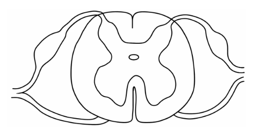
Copy the diagram given below. (a) Name the structure. (b) Label Gray matter and White matter.
Topic: Spinal cord — grey and white matter
Answer
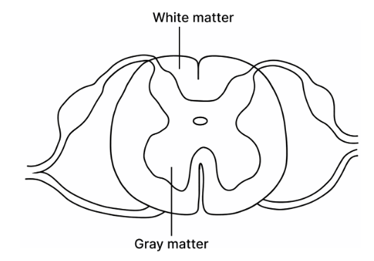
(a) The structure is a TRANSVERSE SECTION (cross-section) OF THE SPINAL CORD.
(b) In your copied diagram: • GREY MATTER — the butterfly-shaped or H-shaped region in the CENTRE, containing the cytons. • WHITE MATTER — the region SURROUNDING it on the outside, containing the myelinated axons.
Identify the structure by the shape of the inner region: a BUTTERFLY or letter H in the centre is the signature of a spinal cord section, and nothing else in the syllabus looks like it.
THE ARRANGEMENT, which must be the right way round: in the SPINAL CORD the grey matter is INSIDE and the white matter OUTSIDE. This is the REVERSE of the brain — the same point tested in Question 4(ii) of this paper, which is worth noticing, since the examiner has asked it twice in different forms.
WHY THE COLOURS: grey matter is made of CYTONS (cell bodies), which have no myelin. White matter is made of AXONS, which look white because of their fatty myelin sheaths.
When a question says COPY the diagram, reproduce it neatly in pencil rather than attempting to trace or describe it, and place labels outside with straight ruled lines.