Study the diagram given below and match the structure with its functions:
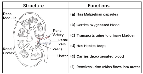
Match each structure of the kidney shown in the diagram with its function: Renal Medulla, Renal Cortex, Renal Artery, Renal Vein, Pelvis, Ureter (a) Has Malpighian capsules (b) Carries oxygenated blood (c) Transports urine to the urinary bladder (d) Has Henle’s loops (e) Carries deoxygenated blood (f) Receives urine which flows into the ureter
Topic: Structure of the kidney
Answer
Renal Medulla – (d) Has Henle’s loops Renal Cortex – (a) Has Malpighian capsules Renal Artery – (b) Carries oxygenated blood Renal Vein – (e) Carries deoxygenated blood Pelvis – (f) Receives urine which flows into the ureter Ureter – (c) Transports urine to the urinary bladder
Work outward from the middle of the kidney and the matching falls into place.
THE TWO REGIONS — and this is the pair most often swapped: the CORTEX is the OUTER region and holds the MALPIGHIAN CAPSULES (Bowman’s capsule with its glomerulus), where filtration happens. the MEDULLA is the INNER region and holds the LOOPS OF HENLE, which run down into it and are responsible for concentrating the urine. Fix it by the logic: filtration must happen where the blood arrives, at the outside; concentration happens as the filtrate travels inwards.
THE TWO VESSELS: the renal ARTERY brings OXYGENATED blood in, the renal VEIN takes DEOXYGENATED blood away. This follows the general rule that arteries carry oxygenated blood and veins deoxygenated — the pulmonary vessels being the one exception in the body.
THE URINE PATH: collecting ducts → PELVIS (the funnel-shaped cavity that collects the urine) → URETER (the tube carrying it to the bladder). Pelvis collects, ureter transports.
In a matching question, do the pairs you are certain of first and let elimination settle the rest.