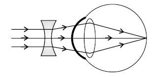
The diagram given below depicts a defect of the human eye which has been corrected by using a suitable lens. Answer the following questions: The diagram given below depicts a defect of the human eye which has been corrected by using a suitable lens. Answer the following questions:
Name the three concentric layers of the eyeball.
Topic: Layers of the eyeball
Answer
From outside inwards:
- SCLERA (sclerotic layer) — the tough protective outer coat
- CHOROID — the middle, vascular and pigmented layer
- RETINA — the innermost, light-sensitive layer
Give them IN ORDER from outside inwards, and add what each does — it costs a few words and shows you know why they differ.
SCLERA — protective. Its transparent front portion is the CORNEA. CHOROID — richly supplied with blood vessels, nourishing the eye, and darkly pigmented to prevent internal reflection of light. It continues in front as the CILIARY BODY and the IRIS. RETINA — contains the RODS and CONES, where the image is formed.
Noting that the cornea, ciliary body and iris are CONTINUATIONS of the outer two layers ties the whole structure together and answers several other questions at once — including the 2025 paper’s question on the ciliary body.