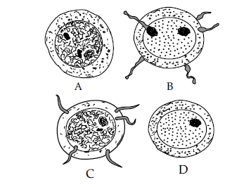
Given below are diagrams showing the different stages in the process of fertilisation of an egg in the human female reproductive tract. Study the diagrams and answer the questions: Given below are diagrams showing the different stages in the process of fertilisation of an egg in the human female reproductive tract. Study the diagrams and answer the questions:
Draw a neat, labelled diagram of a mature human sperm.
Topic: Human sperm — diagram
Answer
Your diagram must show three regions: • HEAD — with the ACROSOME as a cap at the front and the NUCLEUS (containing the chromosomal material) filling most of it, bounded by the PLASMA MEMBRANE • NECK — the short region joining head to middle piece • MIDDLE PIECE — containing MITOCHONDRIA arranged spirally • TAIL — a long whip-like flagellum
Labels required: plasma membrane, acrosome, nucleus, head, neck, middle piece, mitochondria, tail.
THE STRUCTURE-FUNCTION LINK is what the diagram really tests, and one phrase beside each label secures the marks: HEAD — carries the genetic material (23 chromosomes), with the ACROSOME holding HYALURONIDASE, the enzyme that dissolves the covering of the ovum so the sperm can enter MIDDLE PIECE — packed with MITOCHONDRIA that respire to supply the ATP for swimming; the powerhouse of the sperm TAIL — lashes from side to side to propel the sperm forward
So the sperm is stripped to three functions: GENETIC MATERIAL, ENERGY, MOVEMENT. It carries almost no cytoplasm, which keeps it light and fast — exactly the opposite of the OVUM, which is large and full of stored food.
Draw it large and in proportion: the tail should be much the longest part.