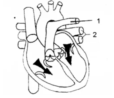
The diagram given below represents a section of the human heart. Answer the questions that follow: The diagram given below represents a section of the human heart. Answer the questions that follow:
Draw neat labelled diagrams of a cross section of an artery and a vein.
Topic: Artery and vein — diagram
Answer
Draw the two side by side, as circles in section.
ARTERY — THICK wall, NARROW lumen, and a circular outline that holds its shape. VEIN — THIN wall, WIDE lumen, and often drawn slightly collapsed or oval. Show a VALVE if the section allows.
Both must show the same three layers, labelled: • TUNICA EXTERNA (outer connective tissue) • TUNICA MEDIA (middle layer of smooth muscle and elastic tissue — drawn THICK in the artery, THIN in the vein) • TUNICA INTERNA / ENDOTHELIUM (inner lining) • LUMEN (the central cavity — NARROW in the artery, WIDE in the vein)
THE CONTRAST MUST BE VISIBLE IN THE DRAWING ITSELF, not only in the labels. Draw the artery with a conspicuously thick wall and small lumen, the vein with a thin wall and large lumen. Two identical circles with different labels earn very little.
BOTH DIFFERENCES FOLLOW FROM PRESSURE, which is the thing worth understanding rather than memorising: an ARTERY receives blood straight from the ventricle at HIGH pressure, so it needs thick elastic walls to withstand and smooth out the surge. High pressure also means blood cannot flow backwards, so NO valves are needed. a VEIN returns blood at LOW pressure, so thin walls suffice — but low pressure means blood COULD flow backwards, especially against gravity in the legs, so VALVES are essential.
Understand the pressure and you can derive every difference instead of learning a table.